Surgical Anatomy of Liver Vessels
DOI:
https://doi.org/10.52340/9789941873232Abstract
The work "Surgical Anatomy of Liver Vessels" highlights the variability of the shape and position of the liver and gallbladder, as well as the features of branching and the relationship between the portal and hepatic veins. Depending on the branching of the intraorgan vessels, the liver is divided into segments.
The work suggests original incisions of the liver, designed in terms of preventing bleeding, as well as maintaining normal blood circulation in the liver.
Liver surgery, which is so successful in the world today, is based on research that was mainly developed in the 1920s-1950s, and on the basis of which it became possible to perform bloodless (or with minimal blood loss) liver resection. The undisputed leader among the authors of this type of research is the French anatomist and surgeon Claude Couinaud, whose research results are recognized as a guideline in liver surgery. However, Professor Shamshe Kevanishvili is also a worthy representative of that small group of researchers for whom it was equally important to prevent bleeding during liver resection and to maintain a complete blood supply to the remaining part after resection. Taking into account the above, the original rates provided by Professor Kevanishvili are especially important when performing so-called atypical liver resections. In this regard, the presented monograph has not lost its importance and fully retains its relevance.
In addition to the above, the additional portal veins and collateral pathways of the portal vein described by Professor Kevanishvili are still considered to be original contributions to the description of the hepatic vasculature.
Also, a very important fact is confirmed in the monograph that the gallbladder veins establish a connection between the left and right branches of the portal vein and such an anatomical feature may contribute to the metastasis of the tumor from one lobe of the liver to another. This discovery was the basis for the liver resection rule (in case of tumor damage to the organ) proposed by Professor Merab Sareli (Izraelashvili), one of the famous students of Professors Sh. Toidze and Sh. Kevanishvili. It involves prophylactic excision of the gallbladder in order to prevent the spread of metastases.
The presented English translation of the work popularizes the ideas and work of the Georgian scientist, and makes clear to foreign researchers the rich methodological possibilities of research (anatomical dissection, preparation of corrosive specimen, X-ray vasography, dissection under a binocular microscope, etc.) which were actively implemented and used by Georgian morphologists at that time.
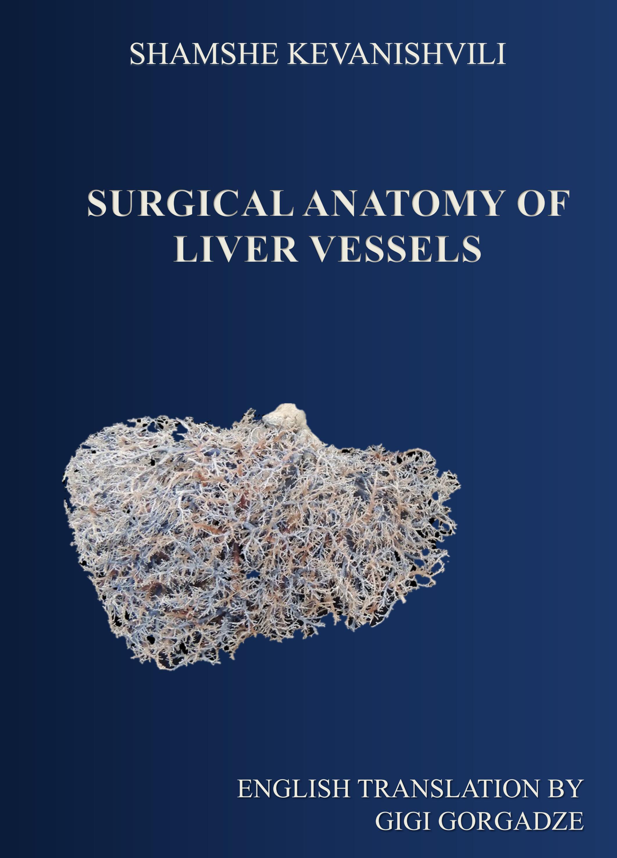
The work is of interest to both morphologists and surgeons. It includes 44 illustrations and 9 tables.
The author of the translation is Gigi Gorgadze, a student of the single-level educational program of medical doctor at Tbilisi State Medical University. The translation was edited by Tamar Turmanidze, Associate Professor of Clinical Anatomy and Operative Surgery Department of TSMU.
Reviewers: Leading Specialist of the Sheba Medical Center - Israel, Foreign Member of the Georgian National Academy of Sciences, Doctor of Medical Sciences, Professor Merab Sareli (Izraelashvili) and Director of TSU Alexander Natishvili Institute of Morphology, Doctor of Medical Sciences, Professor Dimitri Kordzaia.
References
თოიძე შ. თბილ სახ. სამედ. ინსტ. ოპერატიული ქირურგიისა და ტოპოგრაფიული ანატომიის კათედრის შრომები, 1956, I. 89-103.
იოსელიანი დ. თანამედროვე მედიცინა, 1930, II, თბილისი.
კიკნაძე ვ. ღვიძლის ვრცელი რეზექციის გავლენა სისხლწარმოქმნაზე. დის. მედ. მეცნ. დოქტორის ხარისხის მოსაპოვებლად. თბილისი, 1957.
ქევანიშვილი შ. თბილ. სახ. სამედ. ინ-ტის შრომები. ტ. XVIII, 1958, 41-49.
ჯავახიშვილი-კომახიძე ნ. საქ. სსრ მეცნ. აკადემიის ექსპერიმენტული მორფოლოგიის ინსტიტუტის შრომები. ტ. IV, 1953, 65-147.
Акилова А. Т. Труды и материалы Донецкого мединститута, 1936, 1, 20-28.
Альперович Б. И. Вестник хирургии им. И. И. Грекова, 1960, 85, 11, 127-128.
Брегадзе И. Л. Хирургия, № 3, 1957, 26-31.
Бурденко Н. Н. Материалы к вопросу о последствиях перевязки V. Portae. Дисс. Юрьев, 1909.
Валькер Ф. И. К хирургической анатомии системы воротной воны. Дисс. науч. степ. докт. мед. наук, Л., 1920.
Великорецкий А. Н. Хирургия, 1955, 5, 44-54.
Виткинд Ю. Э. Анатомические особенности печени и ее сосудов у детей. Дисс. нз канд. мед. наук, 1936.
Галушко А. Я. Резекция печени в эксперименте и клинике. Дисс. на уч. степ, доктора мед. наук, 1948.
Гудкова Е. С. К анатомин воротной вены. Дисс. на соиск. уч. степ. канд. мед. наук. Горький, 1948. - Делициева К. Н. К типовой печеночных вен. Дисс, на степ. канд. мед. наук. Саратов, 1948.
Делициева К. Н. Труды кафедры нормальной анатомии. Саратов, 1955. 1. 239-243.
Долго-Сабуров Б. А. Об окольном кровотоке в системе воротной вены. Очерки функциональной анатомии кровеносных сосудов. Л., 1961, 148-166.
Золотухин А. Рентгено-ангиология, 1934.
Иоселиани Д. Г. Типовая анатомия брюшной полости новорожденных (в кн. сбори, груд, посвящ. 40 летно научи и учебн. дент. проф. В. Н. Шевкуненко, Л., 1937).
Иоселиани Г. Д. К вопросу патогенеза и лечения симптомокомплекса. Пика. Дисс. на соиск. уч. степ, доктора мед. наук, Тбилиси, 1958.
Касайкина Т. Н. Хирургия, 1953, 7, 62-65.
Ковальский Ф. Ю. О печени у детей. Дисс. СПб, 1900.
Комахидзе М. Э. Тр. ин-та экспе. и клин. хир. и гематологии АН Груз. ССР, т. VII, 1957, 353-372.
Красуская А. А. Двойной ток воротной вены. Известия научного института им. П. Ф. Лесгафта, т. Х, 1924.
Крылова Н. 11. Казанский медицинский журнал, 1960, 4, 63-65.
Кузнецов Б. Г. Ученые записки Горьковского мед. института, 1957, 121-135.
Кулябко Б. В. Изменения в строении в воротной системы печени при нарушениях кровообращения. Л., 1940.
Кунцевич В. В. Труды Военно-мед. Акад. им. С. М. Кирова, том 38, 1947.
Летичевский Б. И. Архив анатомии, гистологии и эмбрио- логин, г. ХХ, 1939.
Лурье А. Вестник хирургии им. И. И. Грекова, 1935, 37, 106 и 107, 187-189.
Максименков А. Н. Крайние типы изменчивости системы нижней полой вены и их прикладное значение. Дис. на уч. степ. докт. мед. наук, Л., 1937.
Мампория Н. М. Микроваскуляризация печени в норме и в эксперименте. Труды Института экспер, морфологии Акад. наук Груз. ССР, т. ІХ, 1961, 95-103.
Мельников А. В. О резекции печени, Хирургия, 1, 1956, 38-17.
Михайлов Г. А. Внутриорганная топография сосудов печени, Ученые записки, т. 111, 1959, 202-209.
Морозова Т. Д. Тезисы докладов II Украинской конференции морфологов, Харьков, 1956.
Надеин А. П. и Крымгольц М. Л. Труды 1 съезда хирургов Закавказья, Баку, 1925.
Надеин А. П. и Крымгольц М. Л. Теорет. и практич.мед.,1926,1,5-6.
Нечунаев Л. М. Казанский медицинский журнал, 1958, 8, 44-51.
Огнев Б. В. и Сызганов А. Н. К хирургической апатомии печени человека, ХVIII съезд Российских хирургов, 1927.
Павлов И. П. Экковский свищ вен нижней полой и воротной и его последствия для организма. Собрание сочинений, М.-Л., 1951, 2, 210-238.
Парфентьева В. Ф. Архитектоника кровеносных сосудовпечени. Кишинев, 1960.
Степанова В. Н. Тр. в/мед. Акад. им. С. М. Кирова, 1953,50,563-579.
Тихомиров М. А. Варианты артерий и вен человеческого тела, Киев, 1899.
Тонков В. Н. Вестник рентгенологии и радиологии, т. ХХѴІ, 5-6, 1947.
Торкачева М. И. Монография, 1924.
Фейтельберг П. И. Анатомия анастомозов между воротной и нижней полой веной в подвздошных областях и по ходу вос ходящего и внсходящего отделов толстых кишок, Дисс. на степ докт. мед. наук, 1947.
Фишман Л. Г. и Кревер А. Н. Вестник рентгенологии и радиалогии, VIII, вып. 1. 1930.
Хашимов Н. Х. Архив анатомии, гистологии и эмбриологии, I 1959, 55-62.
Шевкуненко В. Н. и Максименков А. Н. Новый хир. архив, т. 36, кн. 3-4, 1930.
Шепелев М. В. Вестник хирургии им. И. И. Грекова, 1956, 4, 14-22. Экк Н. В. Военно-медицинский журнал, 1877, ХХХ, 32.
Burlet H. М. Morph. Jahrb, 1911. 42, 1-71.
Couinaud C. Presse méd., 1954, 62, 33, 709-712.
Gans H. Introduction to Hepatic Surgery. 1955.
Glisson F. Anatomia Hepatis. London, 1954.
Hocssetter F. Morph. Jahrb., 1893. 20, 543-648.
Keibel F. U. Mall F. P. Handbuch der Entwicklungsgeschichte des Menschen. Leipzig, 1911, 2.
Kremer K. u. Hilke H. Zentralblatt für Chirurgie, 1959, 31, 1225-1232. Lalanbie. Contribution à Pétude de la circulation intrahè patique. Paris, 1910.
Lewis F. T. Handbuch der Entwicklungsgeschichte des Menchen.
Herausg von Keibel F. und Mall F. P., 1, 911.
Lurie A. Ann. Surg., 1937, 105, 161-168. Referate-Zorg. f. Chir., 83, 144-145.
Melnikoffa. Zeitschrift für Anatomie und Entwicklungsgeschichte, 1294, 70.
Naboer I. F. Frankf. Zeit. Pathol., 1931, 41, 454-11; Refepate-Anat Ber., 25, 462-463.
Perren Tierl. Die Venen der Gallenblase und der extrahepatischen Gallenwege beim Menschen und Wirbeltieren. Stockholm 1933.
Popescu C. Zeitschrift fȃr Arztliche Fortbildung, 1958, 16, 672-676.
Reifferscheid M. Chirurgie der Leber. Klinik und Technik. Stuttgart. 1957.
Ruge G. Leber mit abgespaltenem, rechtem Seitenlappen. Gegenbaurs morphologisches Jahrbuch. 1913, 46, 293.
Sherlock Sc Diseases of the Liver and Biliary System 1958.
Spigel A. Dh humani corporis fabrica 1627.
Downloads
Published
Issue
Section
License

This work is licensed under a Creative Commons Attribution-NonCommercial 4.0 International License.
How to Cite


